

In most fields of immunology, the precise determination of interferon gamma is required. Nevertheless, achieving credible results is not necessarily easy. Variations in sample preparation, inconsistent incubation times, and improper washing can affect assay performance and lead to data that is difficult to interpret.
Consequently, scientists tend to waste more time troubleshooting experiments rather than in data analysis. Fortunately, a user-friendly ELISA workflow can be used to reduce variability and enhance consistency across different experiments.
Every step of the assay adds to the overall accuracy. Thus, it is significant to understand the order of steps to use to obtain reliable outcomes.
Here is a step-by-step guide to using a human interferon gamma ELISA kit, from sample preparation to data analysis.
1. Gather Samples and Assay Components
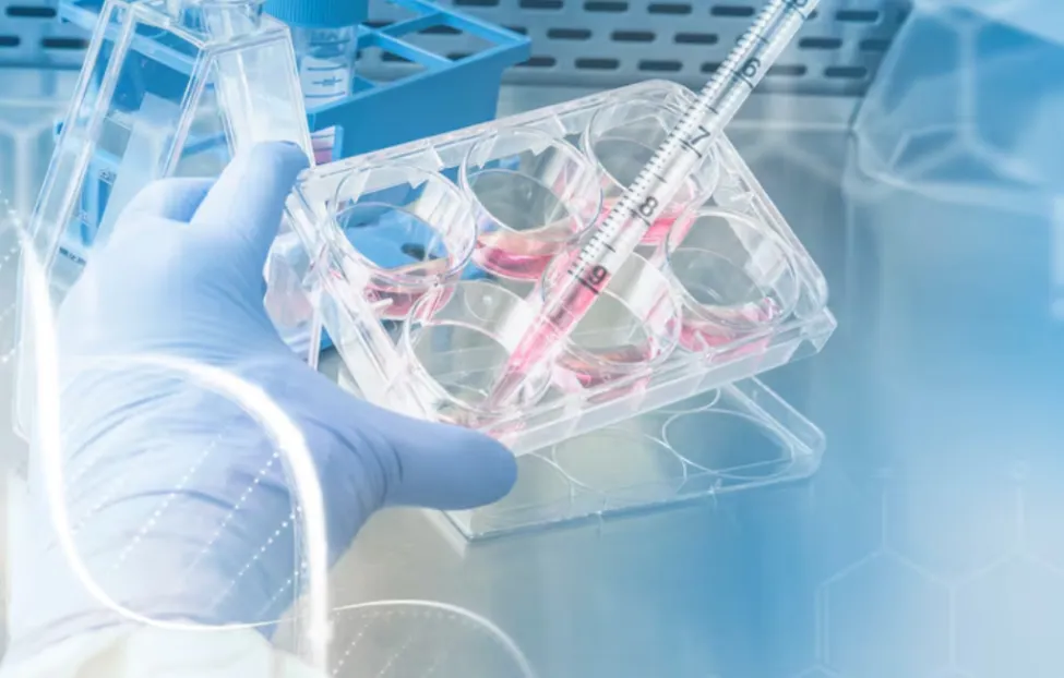
Before starting the assay, make sure all materials are ready for use. The typical contents of a human interferon gamma ELISA kit are a precoated plate, standards, detection antibodies, wash buffer, substrate solution, and other necessary reagents.
Begin by warming all the reagents and samples to room temperature. This assists in keeping the condition of the assays constant. Prepare the wash buffer and any reagent solutions as per the protocol.
Then, prepare your serum, plasma or cell culture supernatant samples. If the samples were kept in the freezer, thaw them and stir them thoroughly. Always avoid repetitive freeze-thaw cycles as they have the potential to interfere with protein stability.
Lastly, label tubes and lay out the plate in advance. Proper preparation eliminates errors and simplifies the rest of the assay processes.
2. Add Standards and Samples to Plate Wells
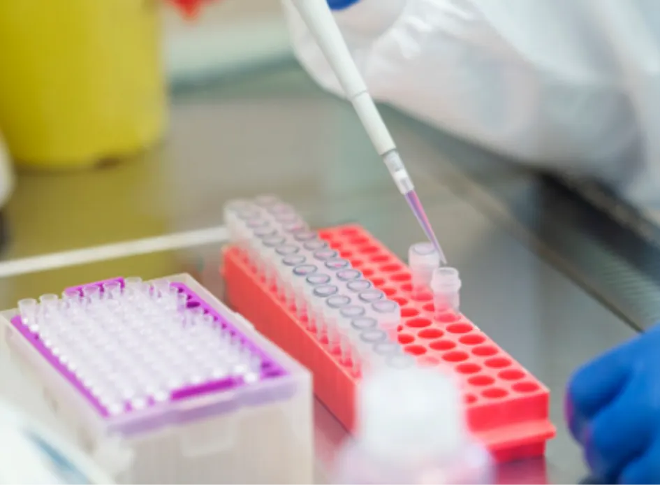
After preparing all reagents and samples, you can begin loading the plate. Begin by putting the standards in their respective wells. These standards have predetermined concentrations of interferon gamma and are employed to form the standard curve in the analysis of the data.
Next, add your samples to the designated wells. Precise transfer of volume with a calibrated pipette. Even small differences in volume can affect the final results.
While pipetting, avoid touching the sides of the wells. This aids in ensuring the plate is in homogeneous conditions. Duplication or triplication of samples is also advisable. Repeated readings assist in enhancing precision and detecting unexpected variations easily.
Plate loading is done carefully in order to achieve reliable protein detection and more consistent assay results.
3. Complete Target Protein Binding Stage
After adding the standards and samples, the next step is incubation. In this step, interferon gamma available in samples attaches to capture antibodies that are coated on the surface of the wells. This interaction is an important part of the sandwich ELISA process.
Adhere to the incubation time and temperature suggested in the assay protocol. Adequate incubation allows the target protein sufficient time to bind efficiently, assisting in enhancing the accuracy of the assays.
Cover the plate during the incubation period. This assists in avoiding contamination and minimizes evaporation, which would influence the results. In addition, ensure that the plate is in a constant temperature environment.
Do not move or shake the plate or disturb it unnecessarily. Stable conditions are used to maintain consistency in all wells. Proper binding on this step helps in the effective detection of the protein and more credible results at later phases of the assay.
4. Apply Detection Antibody and Wash Wells
After the target protein has attached to the capture antibodies, the next step is to add the detection antibody. This antibody attaches to another part of the interferon gamma protein. Together, they form the sandwich complex needed for signal detection.
Once the detection antibody is added, incubate the plate for the time specified in the protocol. This allows proper binding to take place.
After incubation, wash the wells carefully. Washing removes any unbound materials that could interfere with the results. Use the recommended wash buffer and make sure all wells are washed evenly.
In some assays, an enzyme-linked reagent is added after this step, followed by another wash cycle.
Careful washing and consistent handling help reduce background signal and improve the accuracy of interferon gamma measurement.
5. Develop Signal and Measure Absorbance
After the antibody binding steps are finished, the next step is signal development. Begin by adding the substrate solution to each well according to the assay protocol. The enzyme linked to the detection system reacts with the substrate and produces a visible color change inside the wells.
The strength of the color is directly related to the amount of interferon gamma present in the sample. In general, higher protein levels produce a stronger color signal, while lower protein levels produce a lighter signal.
During this stage, it is important to follow the recommended reaction time carefully. If the reaction continues for too long, the signal may become too strong. On the other hand, stopping it too early can reduce sensitivity and affect result accuracy.
Once the development time is complete, add the stop solution to end the reaction. Then place the plate in a microplate reader and measure absorbance at the specified wavelength. Before reading the plate, check each well for bubbles because they can interfere with measurements. Accurate absorbance readings are important for obtaining reliable and reproducible results.
6. Calculate Interferon Gamma Concentration
The final step is to convert absorbance readings into concentration values. First, create a standard curve using the absorbance values from known interferon gamma standards. This curve is used as a reference to find unknown sample concentrations.
Next, compare the absorbance of each sample with the standard curve. You can use analysis software or simple calculations to determine the interferon gamma level in each sample.
After that, check replicate results carefully. Look for any values that are very different from others. Also review control samples to confirm the assay worked properly.
Do not use results that fall outside the valid range of the standard curve. If a sample is too high, dilute it and test again.
Finally, record all results clearly. Good documentation helps keep data accurate and easy to compare in future experiments.
Conclusion
Accurate interferon gamma measurement depends on more than simply following a protocol. Each stage of the ELISA process, from reagent preparation to final data analysis, contributes to the quality and reliability of the results.
Careful sample handling, consistent incubation conditions, effective washing, and precise signal measurement all help reduce variability and improve reproducibility. By following a structured workflow, researchers can generate dependable cytokine data for immunology, inflammation, cell therapy, and related research applications.
A step-by-step approach not only improves assay performance but also helps ensure confidence in experimental findings and long-term study outcomes.
Related posts:
 Voyage Through Time: When will Time Travel be Possible
 Water Damage Restoration Service Lanham: Fast & Efficient
 What Does C3PAO Mean for the Future of Risk Management?
 Creating Stunning Face Swap Videos with AI: How to Use Face Swap Video Tools for Free
 How Dechecker AI Checker Turns Awkward Drafts Into Human-Readable Content
Leave a Reply